Home » Without Label » Upper Back Anatomy - Upper body anatomy, artwork - Stock Image - F006/1233 ... : The bones of the chest and upper back combine to form the strong, protective rib cage around the vital thoracic organs such as the heart and lungs.
Upper Back Anatomy - Upper body anatomy, artwork - Stock Image - F006/1233 ... : The bones of the chest and upper back combine to form the strong, protective rib cage around the vital thoracic organs such as the heart and lungs.
Upper Back Anatomy - Upper body anatomy, artwork - Stock Image - F006/1233 ... : The bones of the chest and upper back combine to form the strong, protective rib cage around the vital thoracic organs such as the heart and lungs.. There is a set of muscles in the upper back (called the thoracic area) called the spinalis thoracis. They originate from the vertebrae and insert into the scapulae. In the upper back region, the trapezius, rhomboid major, and levator scapulae muscles anchor the scapula and clavicle to the spines of several vertebrae and the occipital bone of the skull. Your lower back (lumbar spine) is the anatomic region between your lowest rib and the upper part of the buttock. The trapezius and latissimus dorsi muscles connect the upper limb to the vertebral column.
Covering an expanse from the neck to the tailbone, the back muscles are responsible for a broad range of functions, from extending the spine to shrugging the shoulders. Looking for a solution to your back pain problem? See upper back stock video clips. The cause may be poor posture (such as forward head posture) or any type of irritation of the large back and shoulder muscles, including muscle strain or spasms. The iliocostalis muscles are furthest from the spine.
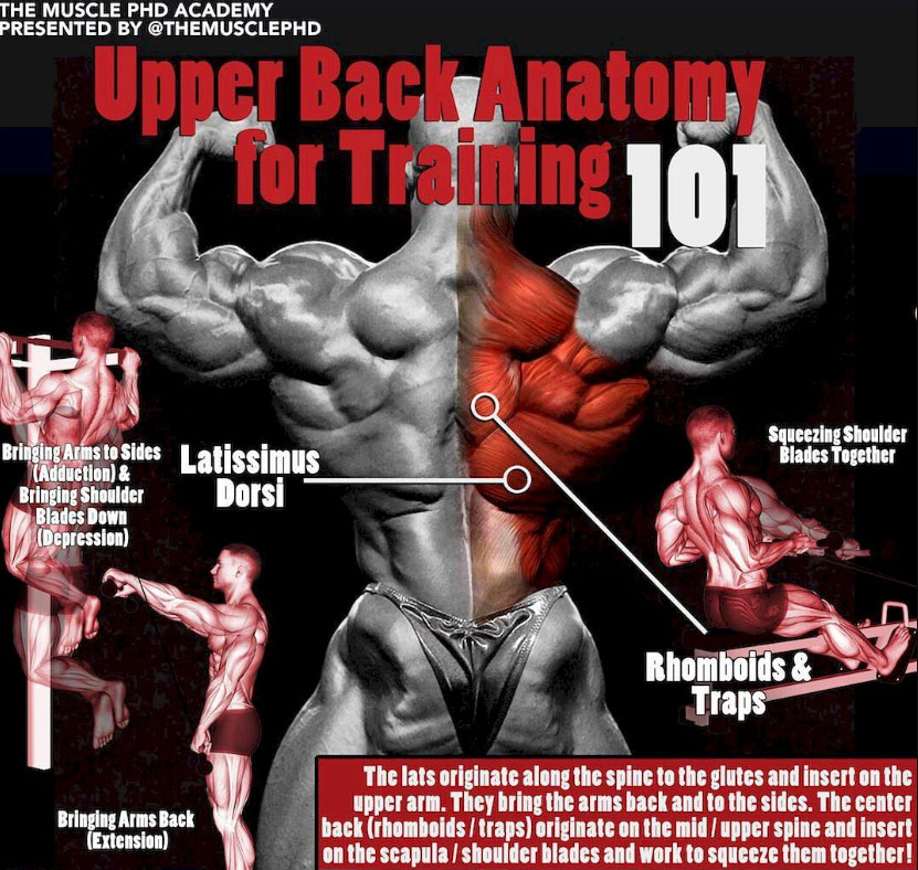
Upper Back Anatomy For Training | Photo & Guide from weighteasyloss.com They originate from the vertebrae and insert into the scapulae. The cause may be poor posture (such as forward head posture) or any type of irritation of the large back and shoulder muscles, including muscle strain or spasms. Your lower back (lumbar spine) is the anatomic region between your lowest rib and the upper part of the buttock. The trapezius and latissimus dorsi muscles connect the upper limb to the vertebral column. The upper back is a complex area containing a number of muscles that perform various actions on the scapulae (shoulder blades) and humerus. There is a set of muscles in the upper back (called the thoracic area) called the spinalis thoracis. It runs from the neck to the upper back. The iliocostalis muscles are furthest from the spine.
The anatomy of the back refers to the muscles of the back, as well as the bones of the scapulae, ribcage, and spine.
The extrinsic (superficial) back muscles, which lie most superficially on the back. The upper back originates at the base of your neck, incorporates both shoulders and extends down to mid spine, including your ribs. There is a set of muscles in the upper back (called the thoracic area) called the spinalis thoracis. The muscles of the chest and upper back occupy the thoracic region of the body inferior to the neck and superior to the abdominal region and include the muscles of the shoulders. The back functions are many, such as to house and protect the spinal cord, hold the body and head upright, and adjust the movements of the upper and lower limbs. The cervical spine is the top part of the spine. The thigh bears much of the load of the body's weight when a person is upright. Powerful muscles that move the head and arms attach to these bones as well. Sometimes you feel the effects right away. This curve, called lordosis, helps to: They originate from the vertebrae and insert into the scapulae. The trapezius and latissimus dorsi muscles connect the upper limb to the vertebral column. Anatomy of the upper back.
The back functions are many, such as to house and protect the spinal cord, hold the body and head upright, and adjust the movements of the upper and lower limbs. Powerful muscles that move the head and arms attach to these bones as well. This is my video about the muscles of the back. This curve, called lordosis, helps to: The back anatomy includes the latissimus dorsi trapezius erector spinae rhomboid teres major.
3D Illustration Back Upper Leg Human Anatomy Stock ... from thumbs.dreamstime.com The back is found posteriorly and includes the vertebral column, the muscles that support the back and the spinal cord. The extrinsic (superficial) back muscles, which lie most superficially on the back. The back muscles are divided into two large groups: 1 your spine in this region has a natural inward curve. These layers of back muscles help to mobilize and stabilize your trunk during your day to day activities. The superficial back muscles are situated underneath the skin and superficial fascia. The rhomboid muscle is activated as you bring and squeeze your scapula or shoulder blades back and together. Both the deltoid and the trapezius are firmly attached to the spine of the scapula.
The bones of the chest and upper back combine to form the strong, protective rib cage around the vital thoracic organs such as the heart and lungs.
It runs from the neck to the upper back. The rhomboid muscle is activated as you bring and squeeze your scapula or shoulder blades back and together. The cervical spine supports the weight and movement of your head and protects the nerves exiting your brain. It comprises the vertebral column (spine) and two compartments of back muscles; In the upper back region, the trapezius, rhomboid major, and levator scapulae muscles anchor the scapula and clavicle to the spines of several vertebrae and the occipital bone of the skull. Both the deltoid and the trapezius are firmly attached to the spine of the scapula. Upper back pain is most commonly caused by muscle irritation or tension, also called myofascial pain. The deltoid, teres major, teres minor, infraspinatus, supraspinatus (not shown) and subscapularis muscles (not shown) all extend from the scapula to the humerus and act on the shoulder joint. The thigh bears much of the load of the body's weight when a person is upright. The hurt can stem from sore muscles, ligaments, and tendons, or from herniated disks, fractures, and other problems in your upper, middle, and lower back. Try the injurymap exercise app now. The cervical spine protects the nerves connecting to. Your lower back (lumbar spine) is the anatomic region between your lowest rib and the upper part of the buttock.
It contains many muscles and nerves but only has one bone, the femur, which is the longest and strongest bone in. Anatomy of the upper back. The upper back originates at the base of your neck, incorporates both shoulders and extends down to mid spine, including your ribs. The upper back is a complex area containing a number of muscles that perform various actions on the scapulae (shoulder blades) and humerus. These layers of back muscles help to mobilize and stabilize your trunk during your day to day activities.
Upper Back Anatomy Muscles : Deep Muscles of the Back ... from s-media-cache-ak0.pinimg.com Looking for a solution to your back pain problem? Related posts of upper back muscle diagram muscle anatomy diagram. The cervical spine supports the weight and movement of your head and protects the nerves exiting your brain. Powerful muscles that move the head and arms attach to these bones as well. The teres major muscle originates on the outer (lateral) edge of the scapula and attaches to the humerus. The anatomy of the back refers to the muscles of the back, as well as the bones of the scapulae, ribcage, and spine. Back muscles anatomy here include the trapezius, latissimus dorsi, rhomboid and levator scapulae. The thigh bears much of the load of the body's weight when a person is upright.
All these muscles are therefore associated with movements of the upper limb.
The upper back originates at the base of your neck, incorporates both shoulders and extends down to mid spine, including your ribs. All these muscles are therefore associated with movements of the upper limb. There is a set of muscles in the upper back (called the thoracic area) called the spinalis thoracis. The bones of the chest and upper back combine to form the strong, protective rib cage around the vital thoracic organs such as the heart and lungs. These muscles are also called immigrant muscles, since they actually represent muscles of the upper limb that have migrated to the back during fetal development. The upper back is a complex area containing a number of muscles that perform various actions on the scapulae (shoulder blades) and humerus. The rib cage also anchors the bones of the head, neck, shoulders, and arms to the trunk of the body. Try the injurymap exercise app now. Related posts of upper back muscle diagram muscle anatomy diagram. The cervical spine protects the nerves connecting to. The cervical spine supports the weight and movement of your head and protects the nerves exiting your brain. The basic anatomy of your upper back by lindsey mcfadden as you're doing your regular upper back stretching exercises , you're probably wondering about the components of your upper back and why it happens to be the most stable part of your spine. The extrinsic (superficial) back muscles, which lie most superficially on the back.