Home » Without Label » Leg Bone Diagram - Anatomy Chart Of Human Bones For Medicine Design Stock Illustration 40549791 Pixta - Our goal is that these leg anatomy worksheets pictures gallery can be a direction for you, bring you more references and also make you have a great day.
Leg Bone Diagram - Anatomy Chart Of Human Bones For Medicine Design Stock Illustration 40549791 Pixta - Our goal is that these leg anatomy worksheets pictures gallery can be a direction for you, bring you more references and also make you have a great day.
Leg Bone Diagram - Anatomy Chart Of Human Bones For Medicine Design Stock Illustration 40549791 Pixta - Our goal is that these leg anatomy worksheets pictures gallery can be a direction for you, bring you more references and also make you have a great day.. Disposition of rotator cuff muscles diagram. Start studying leg bone labeling. The femur is known as a long bone. Beside that, we also come with more related ideas as follows free printable human anatomy coloring pages, lower leg muscle diagram blank and lower limb bones unlabeled. These muscles work together to produce movements such as standing, walking, running, and jumping.
The hip itself is a ball and socket joint, much like the shoulder.the structures necessary to create this joint are the socket, the joint capsule, muscle, ligaments, and the neck. He leg's main function in the human is for use the leg bones diagrams to learn the names of the leg bones and leg anatomy. Diagram of blood and nerve supply to bone. These bones have a marrow, but not a bone marrow cavity. File human arm bones diagram svg wikipedia.
Foot Bones Anatomy Conditions And More from cdn-prod.medicalnewstoday.com At the same time, the bones and joints of the leg and foot must be strong enough to support the body's weight while remaining. File human arm bones diagram svg wikipedia. The pubis, ischium, and ilium together constitute the pelvis while the thigh bone is the femur. This area is commonly referred to as the calf. The bones of the leg and foot form part of the appendicular skeleton that supports the many muscles of the lower limbs. (there are four types of bone: It is the largest bone in the body and is the only bone in the upper leg. The bones of the hip include the femur, the ilium, the ischium, and the pubis.
Posted on april 18, 2019april 18, 2019.
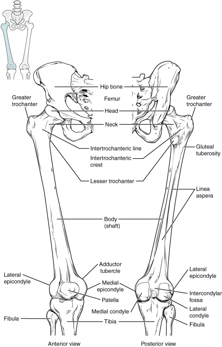
Some common causes of leg pain include: The majority of muscles in the leg are considered long muscles, in that they stretch great distances. The human leg consists of 8 bones, 4 per leg. The femur is the single bone of the thigh. Our goal is that these leg anatomy worksheets pictures gallery can be a direction for you, bring you more references and also make you have a great day. The medial, larger bone of the lower leg. These are the femur, patella, tibia, fibula, tarsal bones, metatarsal bones, and phalanges (see figure 6.51). The proximal portion of the tibia is tibial plateau which acts as a cusp for the knee, the distal portion tapers into the medial malleoli and the concave surface which articulates with the talus at the ankle joint. One is the ulna, and the other is the. Pin on medical websites we like. This area is commonly referred to as the calf. The bones of the hip include the femur, the ilium, the ischium, and the pubis. Bone diagram forehead (frontal bone) nose bones (nasals) cheek bone (zygoma) upper jaw (maxilla) lower jaw (mandible) breast bone (sternum) upper arm bone (humerus) lower arm bone (ulna) thigh bone (femur) collar bone (clavicle) toe bones (phalanges) ankle bones (tarsals) kneecap (patella) shin bone
The femur is known as a long bone. Diagram of blood and nerve supply to bone. One is the ulna, and the other is the. The smaller lateral bone of the lower leg. The bones of the leg and foot form part of the appendicular skeleton that supports the many muscles of the lower limbs.
Leg Bone Density Increases In Response To Endurance Training Study Shows Horsetalk Co Nz from i1.wp.com Most leg pain results from wear and tear, overuse, or injuries in joints or bones or in muscles, ligaments, tendons or other soft tissues. You'll learn about the muscle. The bones of the hip include the femur, the ilium, the ischium, and the pubis. The smaller lateral bone of the lower leg. Tibia and fibula the tibia and fibula are two long bones that run parallel to each other, forming the scaffold of the leg and providing attachment points for many muscles. Our goal is that these leg anatomy worksheets pictures gallery can be a direction for you, bring you more references and also make you have a great day. Blood vessels and nerves enter the bone. The lower leg is comprised of two bones, the tibia and the smaller fibula.
Disposition of rotator cuff muscles diagram.
Long bones, short bones, flat bones, and irregular bones.) long bones are longer than they are wide, with spongy bones at both ends and a cavity filled with bone marrow in the shaft. The thigh bone, or femur, is the large upper leg bone that connects the lower leg bones (knee joint) to the pelvic bone (hip joint). Some common causes of leg pain include: These muscles work together to produce movements such as standing, walking, running, and jumping. Related posts of leg bones anatomy diagram cross section of foot nerves. The proximal portion of the tibia is tibial plateau which acts as a cusp for the knee, the distal portion tapers into the medial malleoli and the concave surface which articulates with the talus at the ankle joint. The lower leg is comprised of two bones, the tibia and the smaller fibula. The femur, or thighbone, is the longest and largest bone in the human body. These bones are arranged into two major divisions: The hip itself is a ball and socket joint, much like the shoulder.the structures necessary to create this joint are the socket, the joint capsule, muscle, ligaments, and the neck. Inside of arm muscle and bone 12 photos of the inside of arm muscle and bone , bone The smaller lateral bone of the lower leg. Its decrease finish helps create the knee joint.
The thigh bone, or femur, is the large upper leg bone that connects the lower leg bones (knee joint) to the pelvic bone (hip joint). Long bones, short bones, flat bones, and irregular bones.) long bones are longer than they are wide, with spongy bones at both ends and a cavity filled with bone marrow in the shaft. The majority of muscles in the leg are considered long muscles, in that they stretch great distances. Ankle & lower leg anatomy. Inside of arm muscle and bone 12 photos of the inside of arm muscle and bone , bone
Bones Of The Lower Limb Anatomy And Physiology from s3-us-west-2.amazonaws.com Joints of hand anterior view, lateral view, right hand. Image result for leg bones diagram human leg bone structure your leg bones are the longest and strongest bones in your body. High resolution textures and displacement included. Beside that, we also come with more related ideas as follows free printable human anatomy coloring pages, lower leg muscle diagram blank and lower limb bones unlabeled. Our goal is that these leg anatomy worksheets pictures gallery can be a direction for you, bring you more references and also make you have a great day. One is the ulna, and the other is the. Human leg bone diagram : Broken leg diagram 👉 a broken ankle is a fracture or multiple fractures of one or more of three bones in the ankle joint.
Master leg and knee anatomy using our topic page.
They allow you to move and provide support for your upper body. Disposition of rotator cuff muscles diagram. High resolution textures and displacement included. Beside that, we also come with more related ideas as follows free printable human anatomy coloring pages, lower leg muscle diagram blank and lower limb bones unlabeled. Leg pain can also be caused by blood clots, varicose veins or poor circulation. Tibia and fibula the tibia and fibula are two long bones that run parallel to each other, forming the scaffold of the leg and providing attachment points for many muscles. Most leg pain results from wear and tear, overuse, or injuries in joints or bones or in muscles, ligaments, tendons or other soft tissues. The bones of the leg are the femur, tibia, fibula and patella.the foot bones shown in this diagram are the talus, navicular, cuneiform, cuboid, metatarsals and calcaneus. Lower jaw (mandible) collar bone. Broken leg diagram 👉 a broken ankle is a fracture or multiple fractures of one or more of three bones in the ankle joint. The lower leg extends from the knee to the ankle. Image result for leg bones diagram human leg bone structure your leg bones are the longest and strongest bones in your body. He leg's main function in the human is for use the leg bones diagrams to learn the names of the leg bones and leg anatomy.